Catalogue

Mouse anti P53 (TP53 tumor suppressor protein)
Catalog number: MUB1501P$422.00
Add To Cart| Clone | DO-1 |
| Isotype | IgG2a/kappa |
| Product Type |
Primary Antibodies |
| Units | 100µg |
| Host | Mouse |
| Species Reactivity |
Human |
| Application |
ELISA Flow Cytometry Immunoblotting Immunocytochemistry Immunofluorescence Immunohistochemistry (frozen) Immunohistochemistry (paraffin) Western Blotting |
Background
P53 is a tumor suppressor gene expressed in a wide variety of tissue types and is involved in regulating cell growth, replication, and apoptosis. It binds to MDM2, SV40 T antigen and human papilloma virus E6 protein. Alterations of the p53 tumor suppressor gene are considered critical events in multistage carcinogenesis of a wide range of human cancers. The mutations found in sporadic cancer are commonly missense mutations that inactivate the tumor suppressor activity of p53. These mutant proteins are much more stable than the normal p53 protein, frequently adopt an altered conformation and accumulate within the nucleus of the malignant cells. Mutations involving p53 are found in a wide variety of malignant tumors, including breast, ovarian, bladder, colon, lung, and melanoma. Positive nuclear staining with p53 antibodies has been reported to be a negative prognostic factor in amongst others breast carcinoma, lung carcinoma, colorectal, and urothelial carcinoma.
Source
DO-1 is a mouse monoclonal IgG2a antibody derived by fusion of mouse myeloma Sp2/0-Ag14 cells with spleen cells from BALB/c mice immunized with recombinant human wild type p53 protein expressed in E. coli.
Product
Each vial contains 100ul 1mg/ml purified monoclonal antibody in phosphate buffered saline (PBS) containing 0.05% sodium azide.
DO-1 recognizes a 53kDa protein, which is identified as p53 suppressor gene product. It reacts with the mutant as well as the wild form of p53. Its epitope maps within the N-terminus (amino acids 11-25) of p53.
Applications
The DO-1 antibody is suitable for the detection of p53 by Western blotting and detects a band of 53kDa in tumor tissue and cell extracts.
The antibody is suitable for immunocytochemistry on permeabilized cells, and Immunohistochemistry on frozen and formalin fixed, paraffin embedded tissues.
Staining of formalin-fixed tissues requires boiling tissue sections in 10mM citrate buffer, pH 6.0, for 10-20 min followed by cooling at RT for 20 minutes.
The antibody can also be applied in ELISA for the quantification of the p53 protein.
Optimal antibody dilutions for the different applications should be determined by titration. Recommended range is 1:500 – 1:1000 for flow cytometry, 1:500-1:2000 for immunohistochemistry, and 1:500 – 1:1000 for immunoblotting applications.
Storage
The antibody is shipped at ambient temperature and may be stored at +4°C. For prolonged storage prepare appropriate aliquots and store at or below -20°C. Prior to use, an aliquot is thawed slowly in the dark at ambient temperature, spun down again and used to prepare working dilutions by adding sterile phosphate buffered saline (PBS, pH 7.2). Repeated thawing and freezing should be avoided. Working dilutions should be stored at +4°C, not refrozen, and preferably used the same day. If a slight precipitation occurs upon storage, this should be removed by centrifugation. It will not affect the performance or the concentration of the product.
Shipping Conditions: Ship at ambient temperature.
Caution
This product is intended FOR RESEARCH USE ONLY, and FOR TESTS IN VITRO, not for use in diagnostic or therapeutic procedures involving humans or animals. It may contain hazardous ingredients. Please refer to the Safety Data Sheets (SDS) for additional information and proper handling procedures. Dispose product remainders according to local regulations.This datasheet is as accurate as reasonably achievable, but our company accepts no liability for any inaccuracies or omissions in this information.
References
1. Vojtĕsek B, Bártek J, Midgley CA, Lane DP.
An immunochemical analysis of the human nuclear phosphoprotein p53. New monoclonal antibodies and epitope mapping using recombinant p53.
J Immunol Methods. 1992;151(1-2):237-44.
2. Vojtĕsek B, Fisher CJ, Barnes DM, Lane DP.
Comparison between p53 staining in tissue sections and p53 proteins levels measured by an ELISA technique. Br J Cancer. 1993;67(6):1254-8.
3. Stephen CW, Helminen P, Lane DP.
Characterisation of epitopes on human p53 using phage-displayed peptide libraries: insights into antibody-peptide interactions.
J Mol Biol. 1995;248(1):58-78.
Protein Reference(s)
Database Name: SwissProt
Accession Number: P04637
Species Accession: Human
Safety Datasheet(s) for this product:
| EA_Sodium Azide |

|
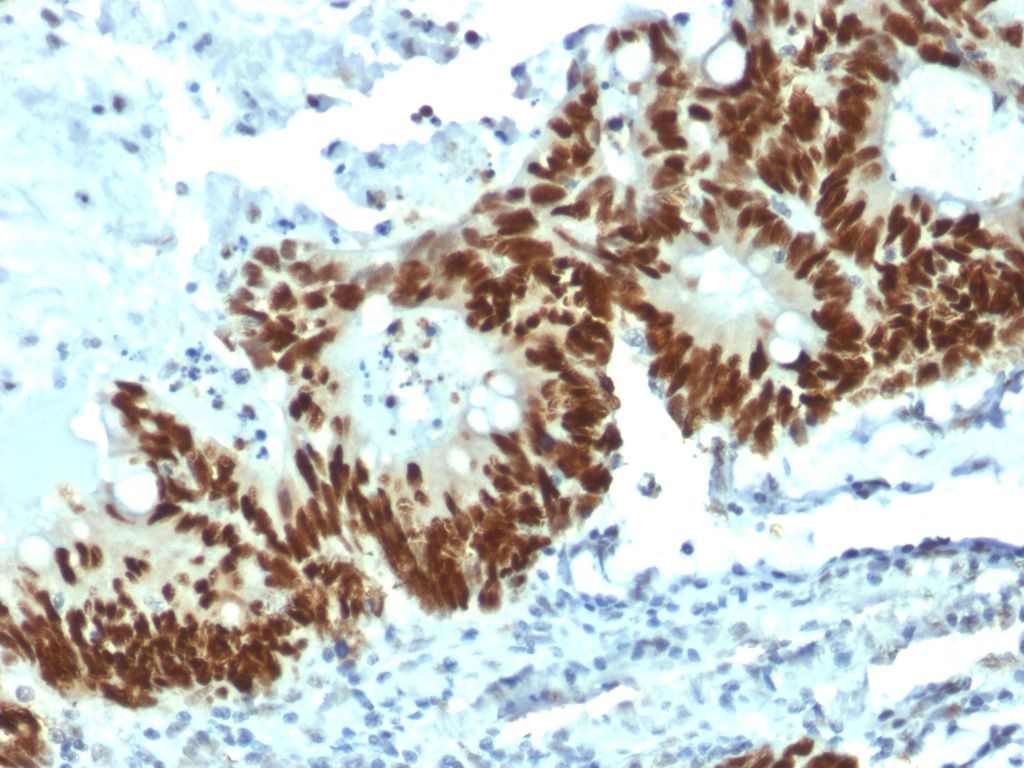
Figure 1: Strong nuclear staining of p53 with MUB1501P (DO-1) in a formalin-fixed, paraffin-embedded human colon carcinoma. |

Figure 1: Strong nuclear staining of p53 with MUB1501P (DO-1) in a formalin-fixed, paraffin-embedded human colon carcinoma.
