Catalogue

Mouse anti Lamin A and C
Catalog number: MUB1102P$422.00
Add To Cart| Clone | 131C3 |
| Isotype | IgG1/kappa |
| Product Type |
Primary Antibodies |
| Units | 0.1 mg |
| Host | Mouse |
| Species Reactivity |
Bovine Canine Hamster Human Mouse Rat Sheep |
| Application |
Flow Cytometry Immunocytochemistry Immunohistochemistry (frozen) Western Blotting |
Background
Nuclear lamins form a network of intermediate-type filaments at the nucleoplasmic site of the nuclear membrane. Two main subtypes of nuclear lamins can be distinguished, i.e. A-type lamins and B-type lamins. The A-type lamins comprise a set of three proteins arising from the same gene by alternative splicing, i.e. lamin A, lamin C and lamin Adel 10, while the B-type lamins include two proteins arising from two distinct genes, i.e. lamin B1 and lamin B2. Recent evidence has revealed that mutations in A-type lamins give rise to a range of rare but dominant genetic disorders, including Emery-Dreifuss muscular dystrophy, dilated cardiomyopathy with conduction-system disease and Dunnigan-type familial partial lipodystrophy. In addition, the expression of A-type lamins coincides with cell differentiation and as A-type lamins specifically interact with chromatin, a role in the regulation of differential gene expression has been suggested for A-type lamins.
Source
131C3 is a Mouse monoclonal IgG1/kappa antibody derived by fusion of P3/X63.Ag8.653 Mouse myeloma cells with spleen cells from a BALB/c Mouse immunized with purified Rat liver lamins.
Product
Each vial contains 100 ul 1 mg/ml purified monoclonal antibody in PBS containing 0.09% sodium azide.
Formulation: Each vial contains 100 ul 1 mg/ml purified monoclonal antibody in PBS containing 0.09% sodium azide.
Specificity
131C3 reacts with an epitope located between residues 319-566 in lamin A and C.
Applications
131C3 is suitable for immunocytochemistry, immunohistochemistry on frozen sections, immunoblotting and flow cytometry. Optimal antibody dilution should be determined by titration; recommended range is 1:100 – 1:200 for flow cytometry, and for immunohistochemistry with avidin-biotinylated Horseradish peroxidase complex (ABC) as detection reagent, and 1:100 – 1:1000 for immunoblotting applications.
Storage
The antibody is shipped at ambient temperature and may be stored at +4°C. For prolonged storage prepare appropriate aliquots and store at or below -20°C. Prior to use, an aliquot is thawed slowly in the dark at ambient temperature, spun down again and used to prepare working dilutions by adding sterile phosphate buffered saline (PBS, pH 7.2). Repeated thawing and freezing should be avoided. Working dilutions should be stored at +4°C, not refrozen, and preferably used the same day. If a slight precipitation occurs upon storage, this should be removed by centrifugation. It will not affect the performance or the concentration of the product.
Shipping Conditions: Ship at ambient temperature.
Caution
This product is intended FOR RESEARCH USE ONLY, and FOR TESTS IN VITRO, not for use in diagnostic or therapeutic procedures involving humans or animals. It may contain hazardous ingredients. Please refer to the Safety Data Sheets (SDS) for additional information and proper handling procedures. Dispose product remainders according to local regulations.This datasheet is as accurate as reasonably achievable, but our company accepts no liability for any inaccuracies or omissions in this information.
References
1. Pugh, G. E., Coates, P. J., Lane, E. B., Raymond, Y., and Quinlan, R. A. (1997). Distinct nuclear assembly pathways for lamins A and C lead to their increase during quiescence in Swiss 3T3 cells, J Cell Sci 110, 2483-93.
2. Neri, L. M., Raymond, Y., Giordano, A., Borgatti, P., Marchisio, M., Capitani, S., and Martelli, A. M. (1999). Spatial distribution of lamin A and B1 in the K562 cell nuclear matrix stabilized with metal ions, J Cell Biochem 75, 36-45.
3. Neri, L. M., Raymond, Y., Giordano, A., Capitani, S., and Martelli, A. M. (1999). Lamin A is part of the internal nucleoskeleton of human erythroleukemia cells, J Cell Physiol 178, 284-95.
Safety Datasheet(s) for this product:
| EA_Sodium Azide |
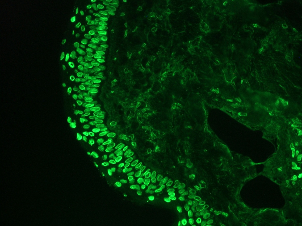
Figure 1. Immunohistochemistry of MUB1102P (131C3) on frozen sections of swine skin showing strong positive staining in nuclei of the epidermal cells and to a lesser extent in the connective tissue. Dilution 1:500.
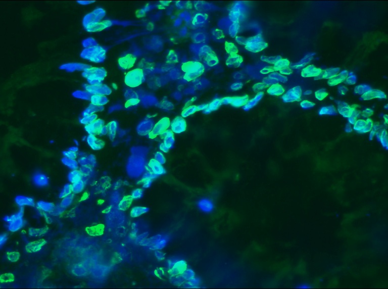
Figure 2. Immunohistochemistry of MUB1102P (131C3) on frozen sections of human colon showing strong positive staining in nuclei of the epithelial cells and to a lesser extent in the connective tissue.
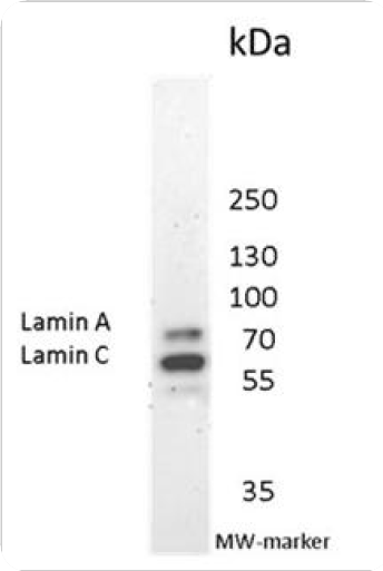
Figure 3. Immunoblotting result of MUB1102P (131C3) recognizing nuclear lamins A and C in human fibroblasts.
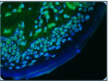
Figure 4. Immunohistochemistry on frozen tissue section of human skin showing nuclear lamina staining in epithelial and connective tissue cells.
