Catalogue

Recombinant Mouse anti Human CD56
Catalog number: 0566P$490.00
Add To Cart| Clone | C5.9 E1 (derived from clone C5.9) |
| Isotype | IgG2b |
| Product Type |
Primary Antibodies |
| Units | 0.1 mg |
| Host | Mouse |
| Species Reactivity |
Human |
| Application |
Flow Cytometry Immunocytochemistry Immunohistochemistry (frozen) Immunohistochemistry (paraffin) |
Background
NCAM / CD56, as a member of the immunoglobulin superfamily of adhesion molecules is characterized by several immunoglobulin (Ig)-like domains. The extracellular part of NCAM consists of five of these Ig domains and two fibronectin type III homology regions. NCAM is encoded by a single copy gene composed of 26 exons. However, at least 20-30 distinct isoforms can be generated by alternative splicing and by posttranslational modifications, such as sialylation. During sialylation, polysialic acid (PSA) carbohydrates are attached to the extracellular part of NCAM. Through its extracellular region, NCAM mediates homophilic interactions. In addition, NCAM can also undergo heterophilic interactions by binding extracellular matrix components, such as laminin, or other cell adhesion molecules, such as integrins.
NCAM can be found in central and peripheral nerve cells, neuroendocrine tissues and at the surface of NK-cells. Also, NCAM is present in malignancies derived from these tissues and cells.
Synonyms: CD56/Neural Cell Adhesion Molecule/NCAM
Source
Mouse monoclonal antibody 0566P is a recombinant CD56 monoclonal antibody produced in mammalian HEK293 cells.
It is produced by cloning the antibody sequence of the mouse anti CD56 hybridoma C5.9, which was derived from the hybridization of mouse Sp2/0 myeloma cells with spleen cells from BALB/c F1 mice immunized with an extract from the KG1a cell line.
Product
Each vial contains 100 ul 1 mg/ml ProtA purified recombinant monoclonal antibody in PBS containing 0.09% sodium azide.
The purity of the antibody is >95% as determined by SDS-Polyacrylamide gel electrophoresis.
The endotoxin level is < 1EU/mg as determined by the LAL chromogenic endotoxin assay.
Purification Method: Protein A/G Chromatography
Concentration: 1 mg/ml
Applications
0566P is suitable for flow cytometry, immunocytochemistry, and immunohistochemistry on frozen and paraffin-embedd tissue sections. Optimal antibody dilution should be determined by titration; recommended range is 1:200 – 1:500 for immunohistochemistry with fluorochrome conjugated secondary antibodies or with avidin-biotinylated horseradish peroxidase complex (ABC) as detection reagent.
Storage
The product should be stored at -20°C. Aliquot to avoid freeze/thaw cycles
Product Stability: See expiration date on vial
Shipping Conditions: The product is shipped at ambient temperature; freeze upon arrival.
Caution
This product is intended FOR RESEARCH USE ONLY, and FOR TESTS IN VITRO, not for use in diagnostic or therapeutic procedures involving humans or animals. It may contain hazardous ingredients. Please refer to the Safety Data Sheets (SDS) for additional information and proper handling procedures. Dispose product remainders according to local regulations.This datasheet is as accurate as reasonably achievable, but our company accepts no liability for any inaccuracies or omissions in this information.
References
1. Hercend T., Griffin J.D., Benussan A., Schmidt R.E., Edson M.A., Brennen A., Murray C., Daley J.F., Schlossman S.F., and Ritz J. Generation of Monoclonal Antibodies to a Human Natural Killer Clone. Characterization of Two Natural Killer-Associated Antigens, NKH1 and NKH2, Expressed on Subset of Large Granular Lymphocytes. J. Clin. Invest. 1985,75: 932.
2. Lanier, L.L., Le, A.M., Civin, C.I., Loken, M.R., and Phillips, J.H. The relationship of CD16 (Leu-11) and Leu-19 (NKH-1) Antigen Expression on Human Peripheral Blood NK Cells and Cytotoxic T Lymphocytes. J Immunol. 1986, 136: 4480.
3. Schmidt RE, Murray C, Daley JF, Schlossman SF, and Ritz J, J. A subset of natural killer cells in peripheral blood displays a mature T cell phenotype. J Exp Med 1986, 164: 351-356
4. Caligiuri M, Murray C, Buchwald D, Levine H, Cheney P, Peterson D, Komaroff AL, and Ritz J. Phenotypic and functional deficiency of natural killer cells in patients with chronic fatigue syndrome. Immunol 1987, 139: 3306-3319
5. Hercend T. and Schmidt R.E. Characteristics and uses of natural killer cells. Immunol Today 1988, 9: 291-293.
6. Muench, Marcus O., et al. Isolation of Definitive Zone and Chromaffin Cells Based upon Expression of CD56 (Neural Cell Adhesion Molecule) in the Human Fetal Adrenal Gland, J Clinical Endocrinology & Metabolism. 2003, 88: 3921-3930,
Protein Reference(s)
Database Name: UniProt
Accession Number: P13591
Species Accession: Human
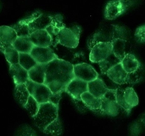
Figure 1. Indirect immunofluorescence staining of human small cell lung cancer cells (NCI-H82) in tissue culture with 0566P (diluted 1:500), showing the specific staining pattern of NCAM/CD56 adhesion sites in between the individual cells.
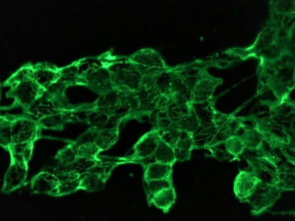
Figure 2. Indirect immunofluorescence staining of human neural cells (SH-SY-5Y) in tissue culture with 0566P (diluted 1:200), showing the specific staining pattern of NCAM/CD56 adhesion sites in between the individual cells.
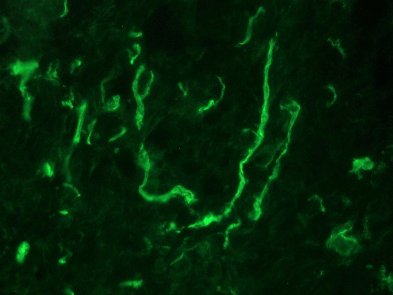
Figure 3. Indirect immunofluorescence staining of human urinary bladder frozen tissue sections with 0566P (diluted 1:500), showing the specific pattern of NCAM/CD56 in the nerves that innervate the tissue. As expected, no reactivity is seen in the epithelial cell compartment, muscle or connective tissue.
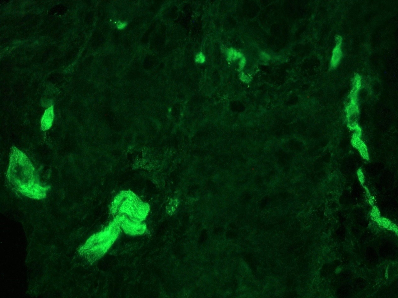
Figure 4. Indirect immunofluorescence staining of swine colon frozen tissue sections with 0566P (diluted 1:500), showing the specific pattern of NCAM/CD56 in the nerves innervating the tissue (parasympathetic ganglion cells in both Auerbach's and Meissner's plexuses). As expected, no reactivity is seen in the epithelial cell compartment, muscle or connective tissue.
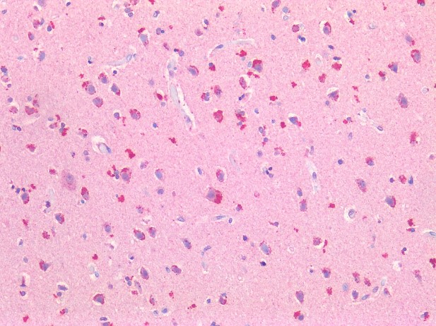
Figure 5. Immunostaining of human formalin fixed, paraffin embedded tissue sections of cerebral cortex with 0566P (diluted 1:400), showing the specific pattern of NCAM/CD56 in the neural cell types. As expected, no reactivity is seen in the connective tissue
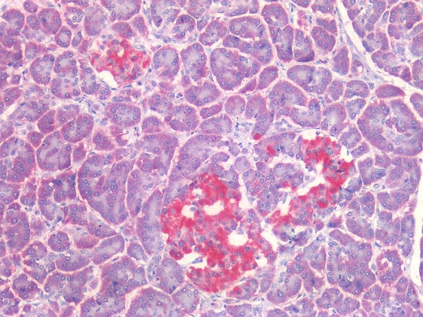
Figure 6. Immunostaining of human formalin fixed, paraffin embedded tissue sections with islets of Langerhans in the pancreas using 0566P (diluted 1:400), showing the specific pattern of NCAM/CD56 in the neuroendocrine cell types. As expected, no reactivity is seen in the epithelial cell compartment or connective tissue
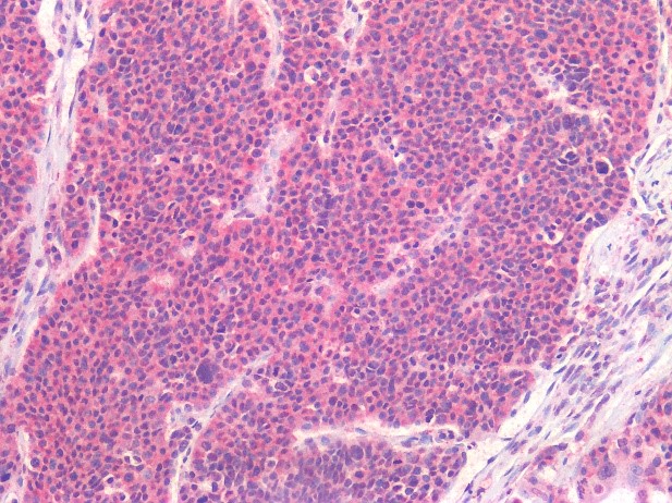
Figure 7. Immunostaining of human formalin fixed, paraffin embedded tissue section of a pancreatic carcinoid with 0566P (diluted 1:400), showing the specific pattern of NCAM/CD56 in the tumor cells.
