Catalogue

Fibronectin, mouse
Catalog number: FI24951$1,050.00
Add To Cart| Product Type |
Polyclonal Antibody |
| Units | 0.5 ml |
| Host | Rabbit |
| Species Reactivity |
Mouse |
| Application |
ELISA Immunofluoresence Immunohistochemistry (frozen & paraffin) Immunohistochemistry (paraffin) Radioimmunoassay |
Background
This antibody may be used to identify mouse fibronectin in experimental studies on wound healing, tumour progression, and tumour invasion, etc. Fibronectins are glycoproteins, which occur as dimers of two 200 kDa subunits. They have binding domains for bacterial proteins, collagens, heparin-like molecules and fibrin. Cellular fibronectin is widely distributed in the stroma of malignant tumours.
Source
Serum from rabbits immunized with purified mouse fibronectin.
Immunogen: Purified fibronectin from murine plasma
Product
Affinity purified (ProtG) antiserum lyophilized from phosphate buffered solution (non-sterile). Contains no BSA or sodium azide! Reconstitute with 0.5 ml of sterile water and store aliquots at -20°C. For further dilution use appropriate antibody diluent, e.g. Art. No. PU002 for IHC.
Purification Method: Protein G affinity purification
Secondary Reagents: Anti-rabbit IgG-conjugates, e.g. anti-rabbit IgG:FITC (Art. No. FI-1000) or anti-rabbit IgG:DyLight488 (Art. No. DI-1488).
Concentration: app. 5 mg/ml
Specificity
Mouse fibronectin
Species Reactivity: Mouse
Applications
IFA, Immunohistochemistry on frozen and paraffin sections, ELISA, RIA
Incubation Time: IHC 60 min at RT or 2-8°C over night
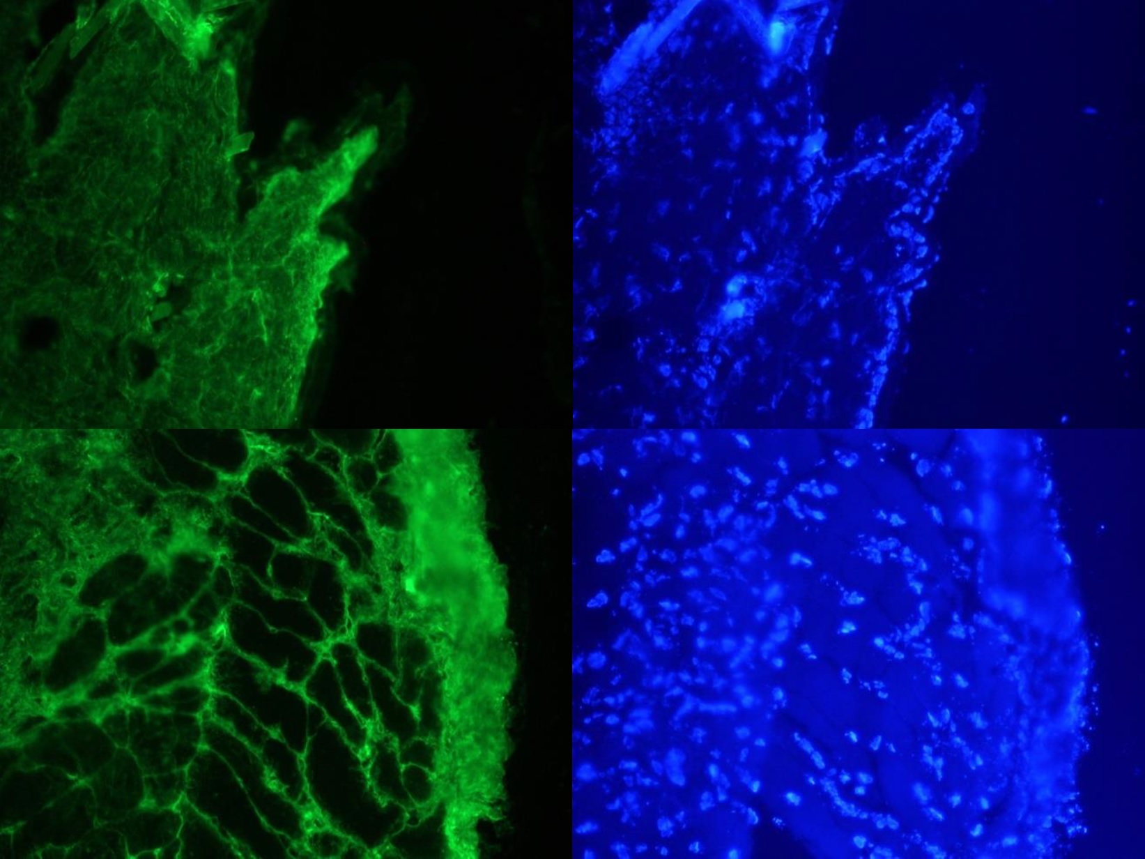
Working Concentration: IHC (frozen): 1:1000, ELISA:1:20000 (OD 0.5) Optimal working dilutions should, however, be determined by antibody titration.
Pre-Treatment: After de-waxing the tissue slices they are treated with 0.2% hyaluronidase (app. 300 U/mg e.g. Art. No. HYA02-50) in TBS 15 min at 37°C. There after non-specific binding is blocked by blocking serum or 3% BSA in TBS. For peroxidase systems blocking with 1% peroxide solution in TBS for 30 min at RT is recommended.
Positive Control: Mouse skin
Storage
-20°C
Caution
This product is intended FOR RESEARCH USE ONLY, and FOR TESTS IN VITRO, not for use in diagnostic or therapeutic procedures involving humans or animals. It may contain hazardous ingredients. Please refer to the Safety Data Sheets (SDS) for additional information and proper handling procedures. Dispose product remainders according to local regulations.This datasheet is as accurate as reasonably achievable, but our company accepts no liability for any inaccuracies or omissions in this information.
References
1. Andujar MB, Magloire H, Hartmann DJ, Ville G, Grimaud JA (1985) Early mouse molar root development: cellular changes and distribution of fibronectin, laminin and type-IV collagen. Differentiation. 30(2):111-22.
2.Savino W, Itoh T, Imhof BA, Dardenne M (1986) Immunohistochemical studies on the phenotype of murine and human thymic stromal cell lines. Thymus. 8(4):245-56.
3. Villa-Verde DM1, Lagrota-Candido JM, Vannier-Santos MA, Chammas R, Brentani RR, Savino W (1994) Extracellular matrix components of the mouse thymus microenvironment. IV. Modulation of thymic nurse cells by extracellular matrix ligands and receptors. Eur J Immunol. 24(3):659-64.
4. Pankov R and Yamada KM (2002) Fibronectin at a glance. J Cell Sci. Oct 15;115(Pt 20):3861-3
5. Labat-Robert J (2002) Fibronectin in malignancy. Semin Cancer Biol. Jun;12(3):187-95.
Protein Reference(s)
Database Name: UniProt
Accession Number: P11276 (FINC_MOUSE)
Species Accession: Mouse
Safety Datasheet(s) for this product:
| EA_Sodium Azide |

Figure 1. Images on the left: Specific indirect immunostaining of connective tissue in frozen sections of mouse skin with FI24951 (diluted 1:1000) Images on the right: Corresponding DAPI staining of nuclei in epidermis, muscle and connective tissue.
